GFC Technique
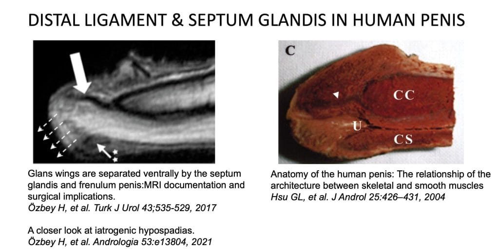
The GFC (Glanular-Frenular Collar) technique is a groundbreaking anatomical approach in modern hypospadias surgery.In hypospadias surgery, tubularization of the neourethra over a catheter or stent and approximation of the dissected glans wings on the midline to enclose the neourethra have been the standard surgical method for decades. However, the male urethra is not a tubular structure with uniform configuration and diameter. The “fossa navicularis” (terminal/glanular portion of the male urethra) has distinct attachments with the “septum glandis” and “frenulum”. Recently, we documented the anatomical features of the glans penis with MRI study. In accordance with previous historical drawings of the glans penis, our findings revealed that a fibrous tissue (septum glandis) covers the glanular urethra (fossa navicularis) circumferentially. It binds the fossa navicularis to the frenulum and corpus cavernosum, as a suspensory ligament on the midline. Hence, it may be true to say that reconstruction of the glanular urethra has been far from its normal anatomical features for decades.
As the most anatomical repair, the GFC technique respects the natural features of the glanular urethra and delivers the most effective and satisfying results in hypospadias.
— Journal of Pediatric Urology, 2017;13:34e1–34e6
The GFC technique is a turning point in hypospadias surgery, which is based on the anatomical features of the penis that are not taken into account since decades. With the GFC technique, the anatomical features of the glanular urethra, in particular the “fossa navicularis”, “septum glandis”, “frenulum” and the “shape of the normal urine stream” are taken into account for the first time. As being the most anatomical repair, the GFC technique gives the most effective and satisfactory results in hypospadias. In normal human penis anatomy, the corpus spongiosum covers the urethra up to the mid-glanular (sub-coronal) level. After that level, a fibrous tissue (septum glandis) surrounds the glanular urethra (fossa navicularis), separates the two hemiglans, connects the upper and lower median septum and holds the glanular urethra in the midline as a suspensory ligament. The glans wings are separated by the ‘septum glandis’ and a ventral cleft between the glans wings that accommodates the frenulum, which is epidermally lined extension of the septum. Hence, the ‘septum glandis’ and frenulum are also included in the formation of the distal (glanular and subcoronal) urethra.
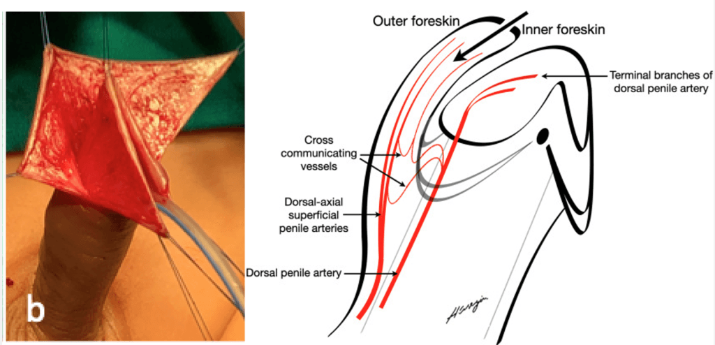
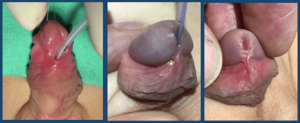
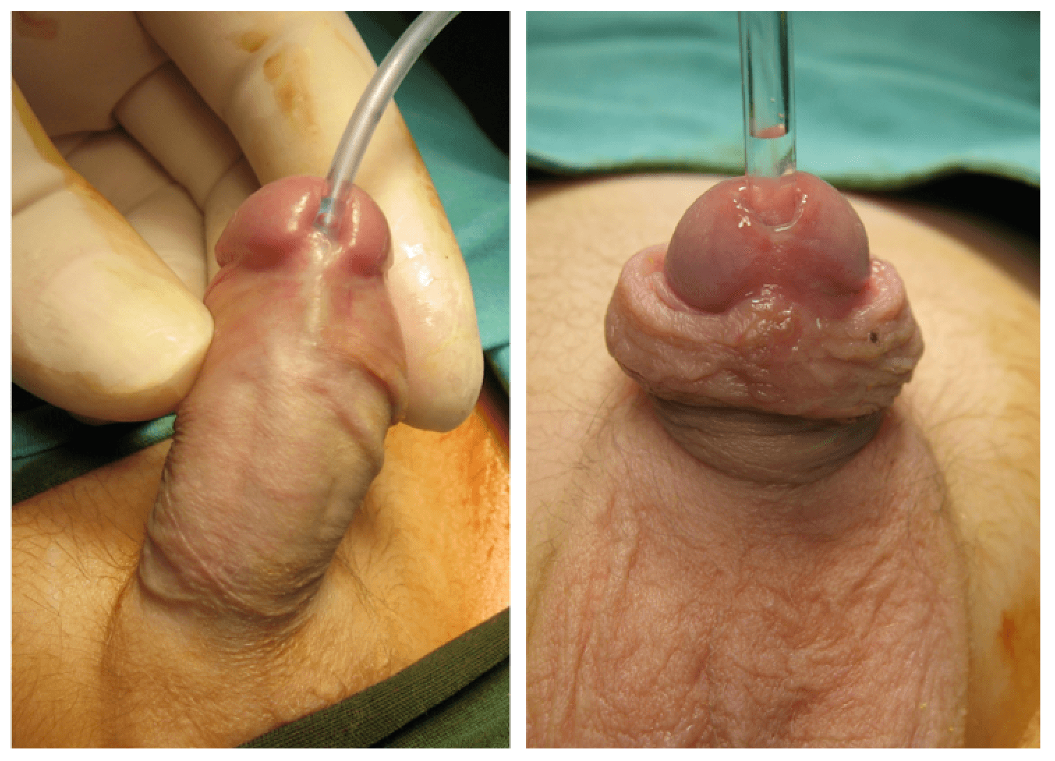
In addition to a defective urethra and its corpus spongiosum, the ‘septum glandis’ and frenulum are entirely missing in hypospadias. The foreskin is not fused ventrally; it appears as a hood over the glans penis. Recent studies have shown that masculinization of the urethral plate occurs in association with the growth and fusion of the preputial fold along the ventral midline of the genital tubercle, which also forms the frenulum of the proximal part of the glanular urethra. Therefore, it is clear that formation of a normal glanular urethra, with its fossa navicularis, ‘septum glandis’ and frenulum are important indicators of an anatomical hypospadias reconstruction. This has inspired a hypospadias repair technique (GFC:Glanular-Frenular Collar technique) that simulates the development of the glanular and subcoronal urethra, which can be incorporated into the repair of all cases of hypospadias. The GFC technique involves reconstruction of the septum glandis, which has been overlooked in the history of hypospadias, and formation of the frenulum, creating the ventral wall of the glanular urethra without dissecting the glans, leaving room for the formation of the navicular fossa. The GFC technique aims to restore the functional anatomy of the glans, replicating the embryologic development of the glanular urethra.
Because of centuries-long misconceptions in repairing the male urethra, hypospadias surgery has become a complex procedure.
— Traditional misconceptions in hypospadias surgery, J Pediatr Surg, 2026
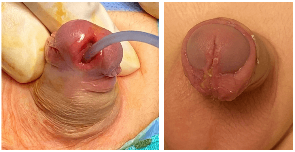
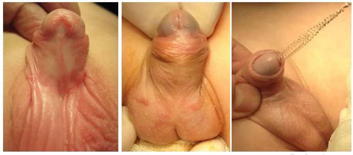
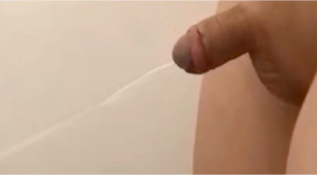
The GFC technique allows a tension-free tubularization of the glanular urethra, afforded by the limited spongioplasty. The space provided for the reformation of the fossa navicularis is supported by loose connective tissue (septum and frenulum) ventrally. The ventral aspect of the glans penis should not be covered (compounded) by the glans wings over its full length, in order to accommodate the frenulum. The GFC technique doesn’t necessitate glans dissection, is neither limited, nor extensive. Subepithelial approximation has been found to be anatomically and physiologically sufficient. With the GFC technique, normal urine flow (wave-like shape) is obtained in all patients. The detailed anatomy of any organ and/or malformation can be further defined based on new knowledge, as in hypospadias. The anatomical differences between the glanular and penile urethra, the structures such as the septum glandis and the fossa navicularis are new definitions that have led to a paradigm shift in hypospadias. The finding that the wings of the glans are not fused together ventrally but are separated by the septum glandis represents a new insight and clarifies the causes of the complications that frequently occur in hypospadias, such as meatal stenosis, fistula and the so-called glans-dehiscence. Reconstruction of hypospadias should include the formation of a septum glandis, frenulum, and navicular fossa with a dual surgical approach for glanular and penile urethra reconstruction.
Hypospadias repair should include reconstruction of the septum glandis, frenulum and fossa navicularis through a dual surgical approach for the glanular and penile urethra.
— The GFC technique: dual approach to hypospadias reconstruction, J Pediatr Urol, 2024
Traditional misconceptions in hypospadias surgery. J Pediatr Surg, January, 2026 (PDF)
The male urethra is not a straight, uniform tube, as reconstructed in all hypospadias repair techniques. It has the “fossa navicularis”, which forms the glanular urethra with specific functional properties. The glans is not an anterior extension or expansion of the corpus spongiosum, which gradually terminates at the mid-glanular level. A faulty development of the fossa navicularis indicates a malformation of the glans itself, manifesting as grooves of varying depth on the glans.
The glans tissue does not completely enclose the navicular fossa, but is separated by the “septum glandis” along the ventral midline. Although there is no “glans fusion”, this has unfortunately been routinely achieved for decades during hypospadias surgery by dissecting the wings of the glans.
The “septum glandis” is the fibroelastic tissue, which covers the fossa navicularis. As a fibroelastic partition, the septum glandis provides the elasticity and adaptive configuration of the glanular urethra during micturition and ejaculation. Together with the frenulum the septum glandis forms the ventral wall of the glanular urethra.
The “distal ligament” contributes significantly to the flexibility and rigidity of the glans, particularly during sexual intercourse. A deep midline incision of the glans is equivalent to a direct visual internal urethrotomy, corresponding exactly to the position of the distal ligament, which has reportedly led to erectile dysfunction in patients following hypospadias repair. Apparently, grafting glans would have far more detrimental consequences.