Home › Hypospadias
What Is Hypospadias? Symptoms and a Comprehensive Treatment Guide
From definition and clinical classification to the ideal age for surgery and home recovery everything families need to know about hypospadias.
Contents

What Is Hypospadias?
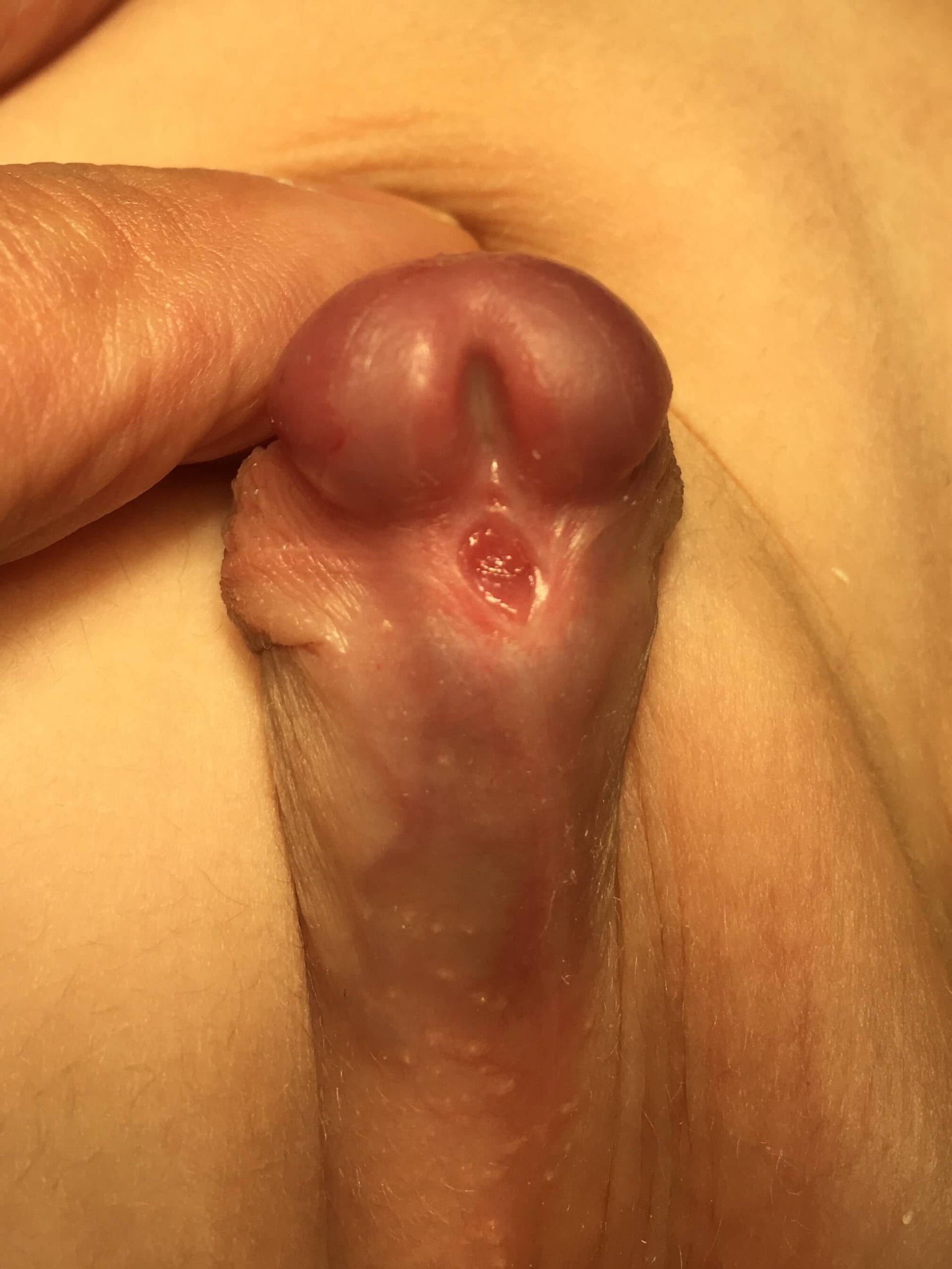
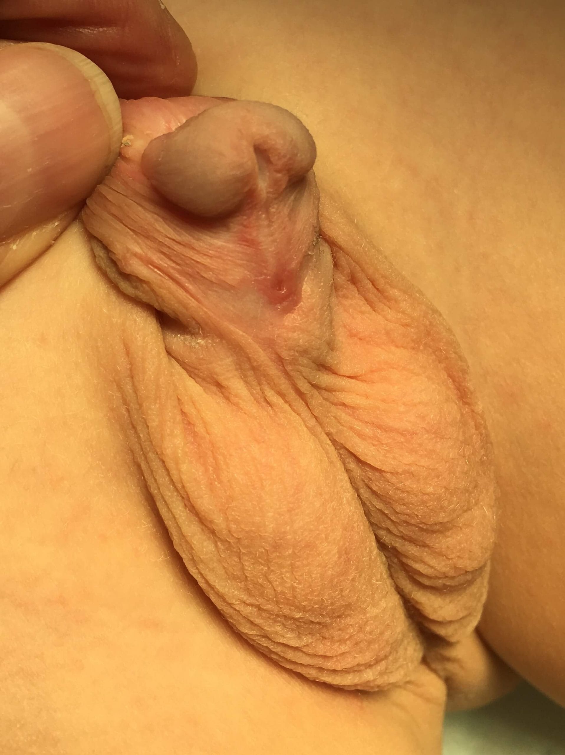
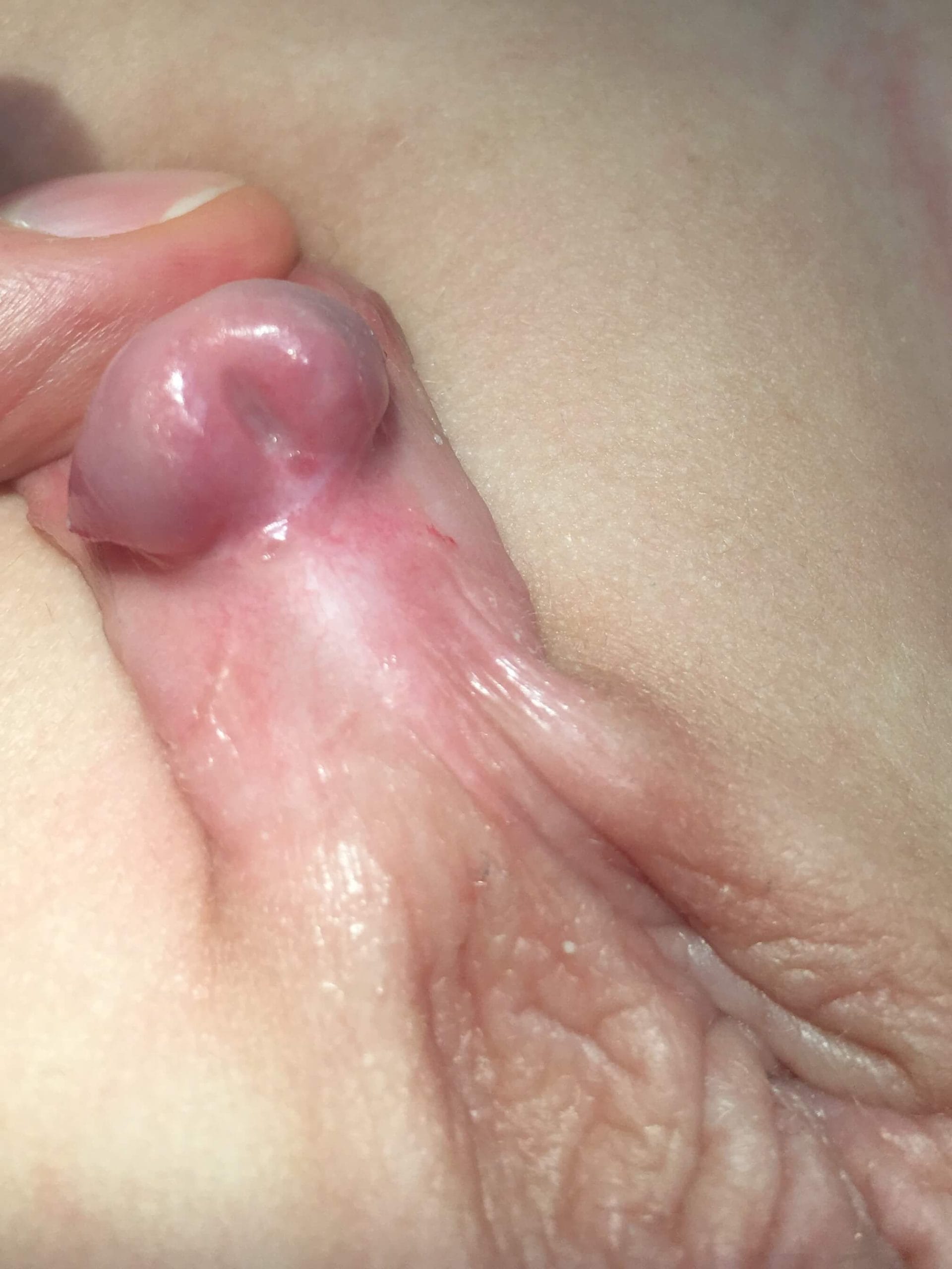
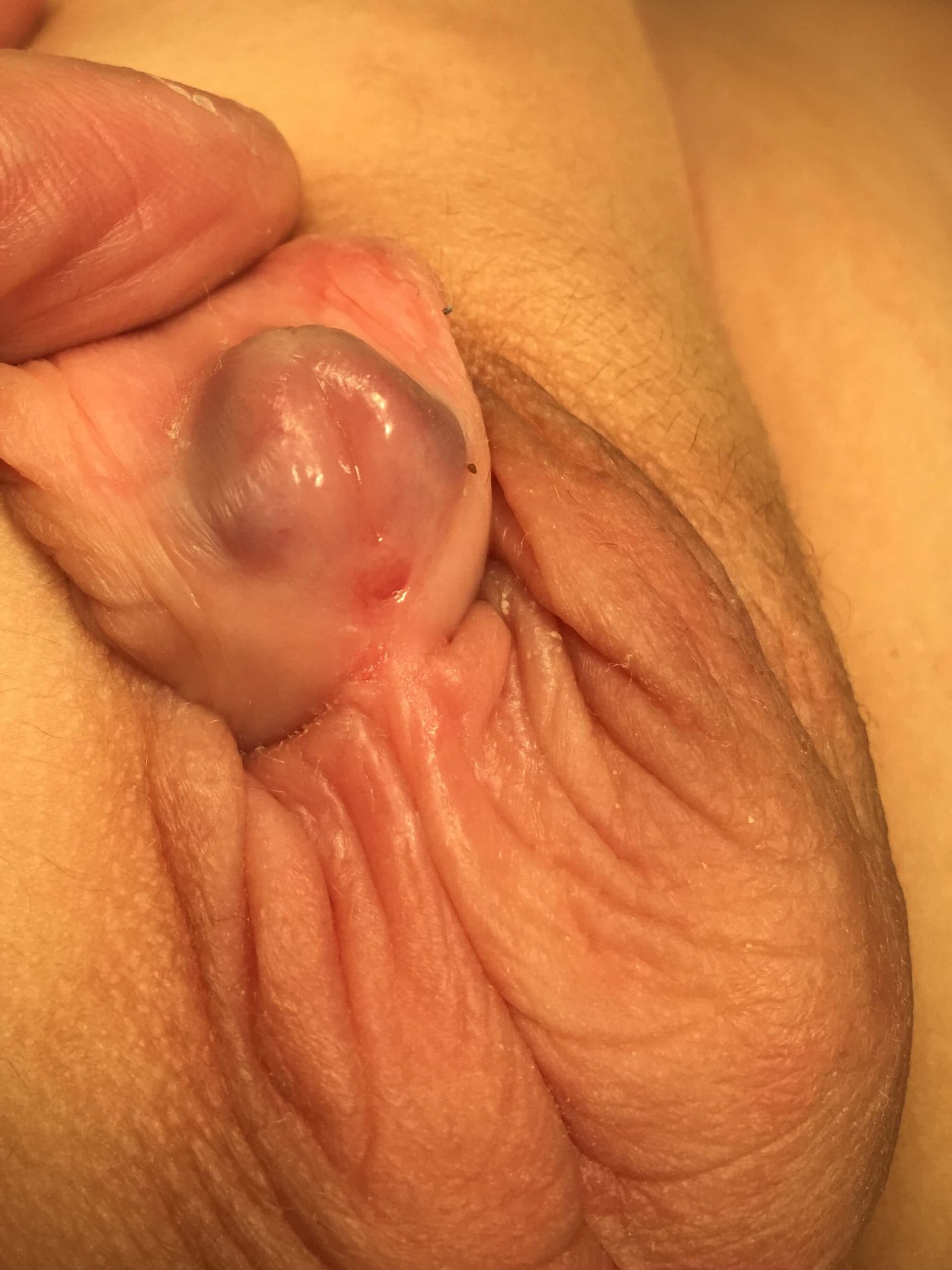
Hypospadias is a congenital anomaly in which the urethral opening (meatus) does not form at the tip of the penis, but instead opens on the underside (ventral aspect) of the glans, along the shaft, or near the scrotum.
Because the foreskin (prepuce) fails to fuse completely on the underside, it gathers excessively on the top (dorsal hood). This characteristic appearance often gives parents the impression that the child was “born half-circumcised.”
It occurs in approximately 1 in 150 to 200 male newborns, making it one of the most common congenital urological conditions. When managed by experienced pediatric urologists and pediatric surgeons, it can be completely and successfully corrected.
Symptoms of Hypospadias: What Should Families Watch For?
The anatomical findings of hypospadias are usually recognized immediately at birth. Clinically, the condition is characterized by four cardinal features:
Ectopic Urethral Opening (Meatus): The opening is located on the underside of the glans, along the penile shaft, near the scrotum, or in the perineal region.
Abnormal Urinary Stream: The urine sprays downward, sideways, or deflects, making it difficult for the child to urinate comfortably while standing as they grow.
Incomplete Prepuce (Dorsal Hood): The foreskin is absent on the underside and bunched over the top of the glans.
Penile Curvature (Chordee): A downward bend of the penis that becomes more distinct during an erection. If left untreated, severe curvature can impact both psychological well-being in childhood and sexual function in adulthood.

Types and Classification of Hypospadias
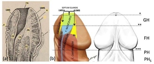
Hypospadias is categorized from mild to severe based on the location of the meatus and the presence of curvature. In addition to defining the anterior urethral anatomy, Prof. Dr. Hüseyin Özbey reclassified hypospadias based on precise anatomical landmarks:
Megameatus Intact Prepuce (MIP): The mildest variant; the foreskin appears intact, but the septum glandis and frenulum are absent, with incomplete formation of the ventral wall of the fossa navicularis.
Glanular Hypospadias (GH – Distal/Mild): The most common form; the meatus is located on the glans, presenting with an underdeveloped, shallow groove at the fossa navicularis.
Frenular Hypospadias (FH – Distal/Moderate): The meatus is located in the frenular zone just beneath the glans, frequently accompanied by glanular tilt.
Penile Hypospadias (PH – Moderate): The meatus opens along the penile shaft, typically associated with downward curvature (chordee).
Proximal Hypospadias (PH-S – Severe): The meatus is situated near the penoscrotal junction or scrotum, accompanied by significant penile curvature that requires comprehensive reconstructive surgery.
When Should Hypospadias Be Treated? (Ideal Age for Surgery)
According to modern pediatric urological guidelines, the optimal window for surgery is between 6 and 18 months of age (up to 2 years). Operating within this period offers distinct advantages:
Psychological Benefit: Surgery is completed before the development of body awareness and gender identity, minimizing psychological trauma.
Rapid Tissue Healing: Infants demonstrate superior tissue regeneration and minimal scarring.
Safe Anesthesia: Modern pediatric anesthesia is exceptionally safe within this age group.
Crucial Rule: Children suspected of having hypospadias must not undergo routine neonatal circumcision, as the dorsal foreskin tissue is vital for surgical reconstruction.

Surgical Repair and Treatment Approaches
The currently accepted standard treatment for hypospadias is surgical reconstruction. The primary objectives of surgery are:
Achieving a smooth, steady, and forward-directed urinary stream with normal caliber and pressure.
Creating an aesthetically natural, slit-like meatus positioned at the tip of the glans.
Ensuring complete correction of penile curvature (orthoplasty) for optimal functional and cosmetic outcomes.
Important Clinical Rule: The foreskin (prepuce) must never be removed (circumcised) before surgery, as it serves as an indispensable source of vascularized tissue for surgical reconstruction.
For this reason, Prof. Dr. Hüseyin Özbey developed the GFC technique, which reconstructs the key anatomical components (septum glandis, fossa navicularis, and frenulum) while fully preserving the natural integrity of the glans.
The GFC Technique in Hypospadias Treatment
Almost all complications in hypospadias treatment stem from a non-anatomical repair of the glanular urethra.
The Glanular-Frenular Collar (GFC) technique is an anatomical breakthrough developed by Prof. Dr. Hüseyin Özbey. Missing structures—the septum glandis, fossa navicularis, and frenulum—are reconstructed without incising or splitting the glans tissue.
By preserving tissue integrity and following the natural embryological anatomy of the urethra, the GFC approach drastically minimizes the risk of fistulas, strictures, and re-operations, delivering superior cosmetic and functional results.
Home Care and Recovery After Hypospadias Surgery
A smooth postoperative recovery relies on simple but essential care principles:
Urethral Stent/Catheter: A small, soft catheter remains in place for several days to direct urine away from the healing tissues.
The “Double-Diaper” Method: Highly recommended to separate bowel movements from the surgical site and keep the catheter secure and clean.
Dressing Care: The surgical bandage protects the delicate tissues and is managed according to the surgeon’s protocol to keep the area clean and dry.
Pain Management: Weight-adjusted pediatric analgesics keep the child comfortable during the first 48–72 hours.
Bathing & Hygiene: Sponge baths are used while the catheter and dressing are in place. Once cleared by the surgeon, the area is gently washed with warm water and patted dry without friction.
Follow-Up Consultations: Scheduled follow-ups ensure that healing, cosmetic alignment, and urinary stream dynamics are progressing as expected.


Frequently Asked Questions (FAQ)
Absolutely not. If hypospadias is diagnosed or suspected, routine circumcision must be strictly avoided. The foreskin (prepuce) is an indispensable source of healthy, vascularized tissue required for the surgical reconstruction of the urethra.
Hypospadias is the second most common congenital urological anomaly in boys, following undescended testes (cryptorchidism). While its global incidence has been steadily rising—with higher rates observed in IVF pregnancies and boys with a family history—the exact etiology is multifactorial:
Endocrine disruptions: Hormonal imbalances affecting androgen synthesis or androgen receptors during fetal development.
Environmental & teratogenic factors: Intrauterine exposure to certain maternal medications or endocrine-disrupting chemicals.
Genetic predispositions: Associated genetic mutations or complex developmental anomalies.
Hypospadias may also present alongside undescended testes, renal anomalies, or disorders of sex development (DSD).
Hypospadias itself does not cause infertility. Once the urethra is reconstructed anatomically and penile curvature is corrected, normal erectile and ejaculatory function is achieved. However, fertility potential may depend on associated conditions—such as undescended testes or hormonal deficiencies—which are evaluated independently.
The duration of the operation typically ranges between 1 and 3 hours, depending on the anatomical severity (distal vs. proximal) and the degree of associated penile curvature (chordee).
In the vast majority of cases, yes. Mild and moderate forms (distal, glanular, and midshaft hypospadias) are routinely corrected in a single-stage procedure. In severe, proximal cases with significant chordee, a staged reconstruction may occasionally be required to ensure the safest long-term outcome.
The majority of postoperative complications—such as meatal stenosis, urethrocutaneous fistula, and glans breakdown—occur because traditional techniques treat the urethra as a simple, straight tube, ignoring the intricate anatomy of the fossa navicularis and the septum glandis. The GFC technique restores these native structures without splitting the glans, significantly reducing complication rates and ensuring a natural, steady urinary stream.
Scientific References
This article was authored and medically reviewed by Prof. Dr. Hüseyin Özbey, Specialist in Pediatric Surgery and Pediatric Urology, based on peer-reviewed medical literature. You can explore his complete list of scientific publications here.
- Özbey H, et al. Hypospadias repair with the GFC Technique. J Pediatr Urol 2017;13:34e1–34e6. DOI
- Özbey H, Kumbasar A. Glans wings are separated ventrally by the septum glandis and frenulum penis: MRI. Turk J Urol 2017;43:525–9. DOI · PMC
- Özbey H, Arlı OT. “Fossa navicularis” and “septum glandis”: a flow-control chamber for the male urethra? Med Hypotheses 2020;140:109642. DOI
- Özbey H. The mystery of Jacob Henle’s “septum glandis”. J Anat 2019;234:728–9. DOI
- Özbey H, Devecioğlu D, Staroverov O. A closer look at iatrogenic hypospadias. Andrologia 2020;53:e13803. DOI
- Özbey H. Anatomical modeling of the foreskin for the reconstruction of glanular hypospadias. J Pediatr Urol 2021;17:335–337. DOI
- Özbey H. The GFC technique: dual approach to hypospadias reconstruction. J Pediatr Urol 2024;20:539–540. DOI
- Özbey H. Boundary tissues and layers in the anatomy of the penis. J Pediatr Urol 2025;21:526–7. DOI
- Özbey H. Traditional misconceptions in hypospadias surgery. J Pediatr Surg 2026. DOI